
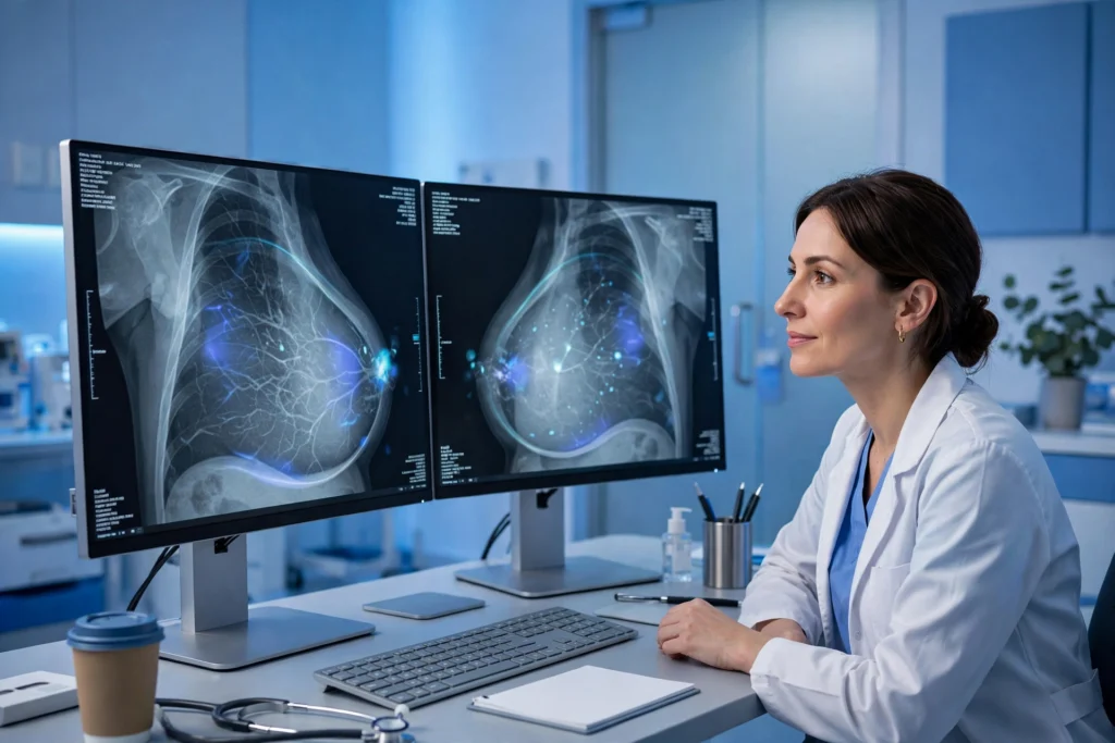
Breast cancer is most treatable when it is found early, often before a lump can be felt or symptoms appear. That simple fact is why screening matters so much — and why a wave of new technology, from artificial intelligence to 3D imaging, is quietly reshaping how the disease is detected. Radiologists still read every mammogram, but increasingly they are not working alone. Software now scans images alongside them, flagging subtle patterns that the human eye can miss and helping clinics decide which patients need a closer look, sooner.
AI as a second pair of eyes
The most visible change is AI-assisted mammography. Trained on millions of earlier scans, modern algorithms can highlight tiny clusters of calcification, asymmetries between the two breasts and changes that have appeared since a woman’s previous appointment. In busy screening programmes, that support matters: research covered by CBS News describes AI tools helping radiologists catch cancers earlier while also reducing the number of women recalled for tests that turn out to be unnecessary. Fewer false alarms mean less anxiety, fewer extra scans and biopsies, and faster answers for everyone.
Doctors are careful to describe the technology as an assistant, not a replacement. An algorithm does not know a patient’s history, symptoms or concerns, and it cannot explain a result or recommend treatment. What it can do is make sure suspicious areas are not overlooked in a long day of reading hundreds of images — the digital equivalent of a diligent colleague double-checking the file before it is signed off.
Better pictures: 3D mammography and beyond
Alongside smarter software, the pictures themselves are improving. Three-dimensional mammography, known as tomosynthesis, takes a series of images from different angles and reconstructs them into layers. Instead of viewing all the tissue flattened into a single picture, radiologists can scroll through the breast slice by slice, which is especially helpful for women with dense breast tissue, where cancers can hide behind overlapping structures on a standard scan.
Other tools are moving from research labs into clinics. Contrast-enhanced mammography uses a dye to highlight areas of increased blood flow, a feature associated with growing tumours. Handheld and automated ultrasound devices are being tested for women whose dense tissue makes mammograms less reliable, and experimental blood tests aim to detect fragments of tumour DNA circulating in the body. None of these replaces regular screening today, but together they point towards detection that is more personalised: the right test, for the right woman, at the right interval.
Risk prediction is getting personal
Technology is also changing who gets screened and how often. Traditional risk calculators lean heavily on age and family history. Newer models combine imaging data, breast density, genetic information and lifestyle factors to estimate an individual’s risk more precisely, so that higher-risk women can be offered earlier or more frequent screening, while lower-risk women may safely avoid unnecessary appointments.
Family history remains a powerful piece of that puzzle, which is why keeping good records matters. If you are organising health documents for yourself or an older relative, our guide to building a family archive starting with what you already have is a practical place to begin — a simple folder noting which relatives had cancer, and at what age, is genuinely useful information for a screening clinic.
What patients should know
First, do not skip screening because the technology is improving. The best scan is the one you actually attend, and national health services continue to recommend regular mammograms for women in the eligible age groups; the National Cancer Institute offers clear background on breast cancer symptoms, screening and treatment. Second, ask questions at your appointment: whether your clinic uses 3D imaging, whether you have dense breasts, and what your results letter actually means. Third, remember that a recall is not a diagnosis — most women called back do not have cancer.
Finally, technology works best alongside awareness. New lumps, changes in shape or skin texture, nipple changes and persistent pain should always be checked promptly, whatever your last screening result said. AI may be changing how breast cancer is found, but the goal is unchanged: find it early, treat it early, and give every patient the best possible chance.
