
Understanding the Condition
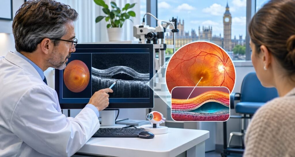
Central serous chorioretinopathy, often shortened to CSCR, happens when fluid collects beneath the macula, the central part of the retina that handles sharp vision. Many people first look for central serous chorioretinopathy treatment in UK clinics after a scan shows this fluid. Understanding what is happening inside the eye makes the next steps easier to follow.
The retina sits at the back of the eye, supported by a pump-like layer of cells and a rich blood vessel layer called the choroid. When either layer misbehaves, fluid can seep into the space under the retina. The cause is not fully understood, and that is why specialists look carefully at each person’s history.
Recognising the Symptoms
Most people notice a painless blur in the middle of their vision, often in one eye. Straight lines such as door frames may look bent. Objects can seem smaller or farther away than normal. Colours may also look dull, and blue shades are often affected first.
The condition most often appears between the ages of 30 and 50, and men are affected more often than women. Reading small print can become tiring. Because the symptoms are painless, some people wait weeks before booking an eye test. An early check helps rule out other macular problems that need different care.
Known Triggers and Risks
Steroid exposure is one of the best-established risk factors. It includes tablets, inhalers, nasal sprays and skin creams. Rarely, a hormone condition that raises the body’s own steroid levels can also be involved. Anyone using steroids should speak with their doctor before changing anything, because stopping some medicines suddenly can be unsafe.
Major stress, disrupted sleep and a driven, competitive personality are also linked with the condition. Researchers are studying genetic differences that may make some people more vulnerable. None of these factors alone explains every case. Doctors therefore review lifestyle, medicines and general health together.
Tests Used After Diagnosis
Optical coherence tomography, or OCT, is the main scan. It uses light waves to create cross-section images of the retina and shows exactly where fluid sits. It takes only minutes, needs no injection and can be repeated to track progress.
Some patients need further imaging. Fluorescein or indocyanine green angiography involves a dye injected into the arm. It highlights leaking spots and helps separate this condition from lookalikes. OCT angiography can check for abnormal new vessels without dye. Your specialist decides which tests are needed, so not everyone has all of them.
Watchful Waiting First
For many people the best first step is careful observation. In roughly 85 per cent of cases, the fluid settles by itself within about six months. During this time, the clinic usually reviews any steroid use and manages other health conditions that might act as triggers.
Regular follow-up scans matter during this period. They confirm the fluid is shrinking and show early if it is not. Patients can support recovery by sleeping well, easing stress where possible and avoiding unnecessary steroid products. If symptoms worsen or flare up repeatedly, the specialist will discuss active treatment.
Photodynamic Therapy Explained
Photodynamic therapy, known as PDT, is widely regarded as a leading option for long-lasting cases. A light-sensitive dye is given through a vein. A low-energy “cold” laser is then directed at the leaking area, which calms the abnormal vessels beneath the retina.
Reports suggest that fluid clears in about 80 per cent of eyes after one or two sessions. Distortion usually improves too. However, vision does not always return fully, even once the fluid has gone. There is a small risk of vision loss, so a specialist will go through suitability and safety before booking treatment.
Laser and Medicine Options
Conventional laser may be considered when the leak is well away from the centre of the macula, because treating close to the centre could damage sight. Micro-pulse laser uses very short, gentle pulses. Recent studies suggest it is less effective than PDT, so it is chosen selectively.
Some tablets have also been studied. Eplerenone and spironolactone, normally used for blood pressure or heart conditions, help certain patients, although results across studies differ. They can alter blood potassium, so blood tests are needed. When abnormal vessels grow under the retina, anti-VEGF eye injections may be used.
Complex Cases and Combinations
Some people have a stubborn or unusual form that does not respond to a single approach. One published clinic example describes a man in his thirties with fibrin-type fluid. He improved partly on a standard anti-VEGF dose, then did better on a higher dose. A combined plan of PDT followed by another injection almost cleared his macular fluid.
Cases like this show why individual planning matters. Specialists at major centres, including London eye hospitals, weigh scan patterns, symptoms and past responses. Anyone looking at central serous chorioretinopathy treatment in UK options should ask whether their case is simple, chronic or complex.
Choosing Care in Britain
Patients can start through an optometrist or GP, who can refer them to hospital eye services. Others choose private retina specialists for quicker appointments and a wider range of scans. Either route should offer OCT imaging and access to PDT or injections when needed.
When comparing providers, ask about experience with this condition, how often they use PDT and how follow-up is arranged. Costs vary, so request a clear written estimate. If you want a second opinion, bring your scan images and a list of every medicine you use. Reliable central serous chorioretinopathy treatment in UK care should always be personal and well explained.
Living and Looking Ahead
The outlook for most people is good, and vision often recovers well. A minority develop long-lasting fluid that can lead to scarring and lasting loss of central vision. This is why symptoms lasting beyond six months deserve a prompt review.
Recurrence can happen, so keep the name of your condition handy and report new distortion quickly. Use an Amsler grid at home if your doctor suggests one. Protect your sleep, manage stress and check with your doctor before using any steroid product. Careful habits and timely appointments give your macula the best chance to heal.
